Sonography
What is Sonography?
Sonography, also known as ultrasound, is a non-invasive diagnostic imaging technique that uses high-frequency sound waves to create real-time images of the internal organs and structures of the body. In gynecology, sonography plays a crucial role in examining the female reproductive organs, including the uterus, ovaries, fallopian tubes, and surrounding tissues. It is commonly used during pregnancy to monitor fetal development and detect any abnormalities. Sonography is a safe and painless procedure that does not involve radiation, making it ideal for diagnosing and monitoring various gynecological and obstetric conditions.
Types of Sonography in Gynecology
There are different types of sonography used in gynecological practice, depending on the area to be examined:
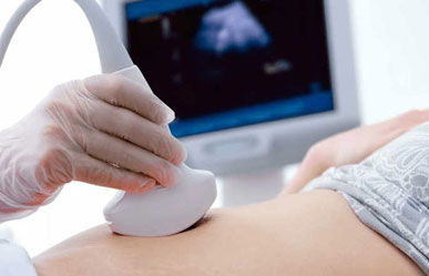
- Abdominal Sonography: This type involves placing the ultrasound probe on the abdomen to obtain images of the uterus, ovaries, and developing fetus during pregnancy. A gel is applied to the abdomen to facilitate sound wave transmission and obtain clear images.
- Transvaginal Sonography (TVS): In this method, a small ultrasound probe is inserted into the vagina to obtain clearer and more detailed images of the uterus, ovaries, and fallopian tubes. It is commonly used to evaluate pelvic pain, abnormal bleeding, infertility, and early pregnancy.
- Doppler Sonography: This specialized form of sonography measures the blood flow in the blood vessels of the uterus, placenta, or developing fetus. It helps in detecting abnormalities such as reduced blood flow to the baby or any placental issues during pregnancy.
Uses of Sonography in Gynecology
Sonography is widely used in gynecology for various diagnostic and monitoring purposes, including:
- Pregnancy Monitoring: Sonography is essential in tracking the growth and development of the fetus, determining the due date, and identifying any potential complications such as ectopic pregnancy, fetal abnormalities, or placenta previa.
- Infertility Evaluation: In cases of infertility, sonography helps assess the condition of the uterus, ovaries, and fallopian tubes to identify any structural abnormalities, cysts, fibroids, or polyps that may affect conception.
- Pelvic Pain and Abnormal Bleeding: Sonography is used to investigate the cause of pelvic pain or abnormal bleeding by examining the uterus, endometrial lining, ovaries, and other pelvic structures.
- Ovarian Cysts and Tumors: Sonography can detect ovarian cysts, tumors, or other abnormal growths in the pelvic region, helping in early diagnosis and treatment.
- Fibroids Detection: Uterine fibroids, which are non-cancerous growths in the uterus, can be diagnosed and monitored through sonography, allowing for appropriate treatment planning.
When to See a Gynaecologist for Sonography?
Women experiencing abnormal menstrual cycles, pelvic pain, difficulty in conceiving, or any pregnancy-related concerns should consult a gynecologist for sonography. Early diagnosis through sonography can help address potential health issues, ensure fetal well-being during pregnancy, and guide the appropriate treatment plan for various gynecological conditions.